
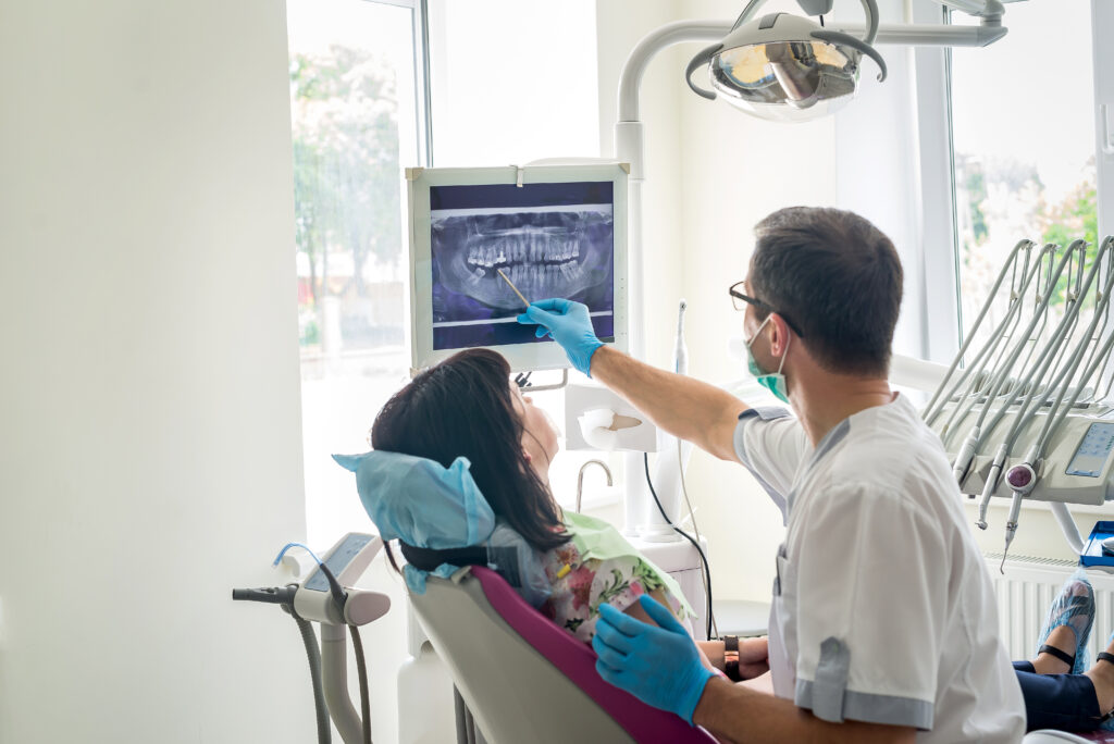
When you settle into the dental chair for a routine check-up, receiving an updated set of dental X-rays, or radiographs, is a fundamental step in your preventative care. While a visual examination allows your dentist to inspect the visible surfaces of your teeth and gums, it only reveals part of the picture. Dental X-rays provide a vital window beneath the surface, allowing your dental team to look inside the teeth and beneath the gumline. By capturing these detailed diagnostic images, potential issues can be detected and intercepted long before they cause discomfort or require extensive, invasive treatment.
The Preventative Power of Diagnostic Imaging
The primary value of routine X-rays lies in their ability to uncover hidden dental developments that are completely invisible to the naked eye. Beneath the surface enamel, small areas of decay can easily develop between adjacent teeth where standard dental mirrors cannot reach. Radiographs also reveal the structural integrity of existing fillings, the early stages of bone loss caused by periodontal disease, and the health of the roots beneath the gums. Identifying these subtle shifts early ensures that treatment can be much more conservative, preserving your natural tooth structure and saving you valuable time and expense over the long term.
Understanding the Safety and Minimal Exposure of Modern Systems
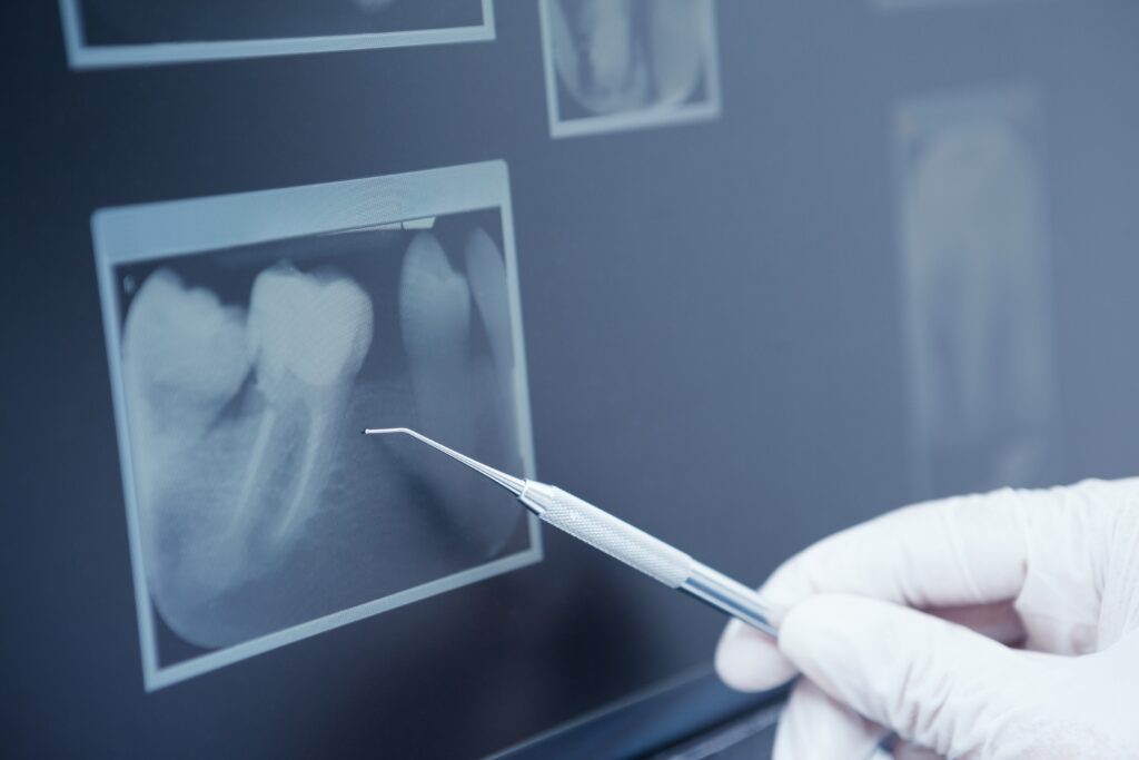
A common question regarding routine radiographs centers on safety and the level of radiation involved. Modern dentistry has experienced significant technological advancements, specifically with the widespread adoption of digital radiography. Digital X-ray systems require a fraction of the radiation exposure used by older, traditional film methods while producing higher-resolution images instantly on a monitor. Furthermore, the administrative use of protective lead aprons and thyroid collars ensures that any secondary exposure is kept virtually non-existent, making the diagnostic benefit of the images far outweigh the minimal risk.
Tailoring Your Frequency to Individual Oral Health Needs
Not every patient requires the exact same sequence or frequency of diagnostic X-rays during their regular visits. For a patient with an exceptionally healthy history, excellent home hygiene habits, and minimal previous dental restorations, a set of bite-wing images may only be necessary every twelve to twenty-four months. Conversely, individuals tracking active decay, undergoing complex structural treatments, or managing ongoing periodontal concerns will benefit from more frequent imaging to closely monitor their progress. Your dental team carefully evaluates your unique oral environment to determine a schedule that safely maximizes your preventative care.
At Bloor Smile Dental in Toronto, the team provides comprehensive family care and patient-focused preventative guidance to help you enjoy a healthy, confident smile. If you want to evaluate your current oral health, require a professional cleaning and examination, or want support maintaining your long-term wellness, you can schedule your next visit with our team today.